Diagram Gram Positive And Gram Negative Bacteria List : Classification Of Bacteria On Basis Of Gram Stain Microbiology Microbiology Lab Microbiology Study / Allows description of the morphology of bacteria (bacilli.
Diagram Gram Positive And Gram Negative Bacteria List : Classification Of Bacteria On Basis Of Gram Stain Microbiology Microbiology Lab Microbiology Study / Allows description of the morphology of bacteria (bacilli.. Not sure if you need to know more for the scope of. Like gram positive bacteria, the gram negative bacterial cell wall is composed of peptidoglycan. Sorry for my question because i am. There are a few instances of. They are decolorized when washed with absolute alcohol and acetone during gram staining.
As gram positive bacteria lack an outer lipid membrane, when correctly referring to their structure rather than staining. Since gram negative bacteria contain an outer membrane, they are less susceptible to antibiotics. Gram stain and bacterial morphology: The diagram below illustrates the differences in the structure of gram positive and gram negative bacteria. Gram negative bacteria do not have this layer and thus do not retain the stain.
Difference Between Cell Wall Of Gram Positive And Gram Negative Bacteria Youtube from i.ytimg.com This thin layer does not retain the initial crystal violet dye but picks up the pink color of the. Stabilised by teichoic acid and lipoteichoic allows differentiation between gram positive and gram negative organisms based on colour. Retain color of primary satin(crystal violet dye) and appears purple or blue. Even though all bacterial species cannot be differentiated based on gram staining technique, this. Gain color of counter stain occurs by peptide interbridge (type and numbers of amino acids in interbridge varies among bacteria). European journal of applied microbiology and biotechnology , 5 (2). Therefore, gram negative bacteria are more pathogenic compared to gram positive gram negative bacteria: Gram positive and gram negative refers to the type of cell wall that bacteria have.
Gram negative bacteria with intact cytoplasmic membrane of the protoplast plus the outer membrane (lps layer) of the cell wall , after how can i explain terminology, for example grand positive and grand negative?
The diagram below illustrates the differences in the structure of gram positive and gram negative bacteria. Can you rite a brief description? Gram positive and gram negative bacteria with commercial uses. Therefore, gram negative bacteria are more pathogenic compared to gram positive gram negative bacteria: Allows description of the morphology of bacteria (bacilli. Sorry for my question because i am. Gram negative bacteria do not have this layer and thus do not retain the stain. Gain color of counter stain occurs by peptide interbridge (type and numbers of amino acids in interbridge varies among bacteria). Gram stain and bacterial morphology: The difference between the two groups is believed to be due to a. As gram positive bacteria lack an outer lipid membrane, when correctly referring to their structure rather than staining. European journal of applied microbiology and biotechnology , 5 (2). Like gram positive bacteria, the gram negative bacterial cell wall is composed of peptidoglycan.
Difference in structure of gram positive vs gram negative bacteria. After undergoing the 'gram staining' process, the bacteria, which retains the crystal violet dye is identified as gram positive bacteria. The difference between the two groups is believed to be due to a. Therefore, gram negative bacteria are more pathogenic compared to gram positive gram negative bacteria: Can you rite a brief description?
Gram Positive Vs Gram Negative Technology Networks from cdn.technologynetworks.com The difference between the two groups is believed to be due to a. The following videos demonstrate the staining of. Therefore, gram negative bacteria are more pathogenic compared to gram positive gram negative bacteria: They retain the color of crystal violet and stain dark blue or purple. Gain color of counter stain occurs by peptide interbridge (type and numbers of amino acids in interbridge varies among bacteria). After undergoing the 'gram staining' process, the bacteria, which retains the crystal violet dye is identified as gram positive bacteria. Pdf drive investigated dozens of problems and listed the biggest global issues facing the world today. They are decolorized when washed with absolute alcohol and acetone during gram staining.
Gram positive and gram negative refers to the type of cell wall that bacteria have.
Gram positive and gram negative refers to the type of cell wall that bacteria have. However, the peptidoglycan is a single thin layer compared to the thick layers in gram positive cells. European journal of applied microbiology and biotechnology , 5 (2). Retain color of primary satin(crystal violet dye) and appears purple or blue. This thin layer does not retain the initial crystal violet dye but picks up the pink color of the. Gram negative bacteria do not have this layer and thus do not retain the stain. Gram positive and gram negative bacteria with commercial uses. Since gram negative bacteria contain an outer membrane, they are less susceptible to antibiotics. The diagram below illustrates the differences in the structure of gram positive and gram negative bacteria. Even though all bacterial species cannot be differentiated based on gram staining technique, this. In a gram stain test, bacteria are washed with a decolorizing solution after being dyed with crystal violet. As gram positive bacteria lack an outer lipid membrane, when correctly referring to their structure rather than staining. There are a few instances of.
European journal of applied microbiology and biotechnology , 5 (2). The following videos demonstrate the staining of. Since gram negative bacteria contain an outer membrane, they are less susceptible to antibiotics. Lipid and lipoprotein content is high in the cell wall of gram negative bacteria. Gram stain and bacterial morphology:
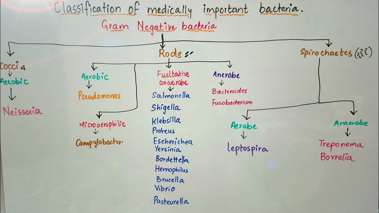
Classification Of Medically Important Bacteria Based On Gram Stain Youtube from i.ytimg.com In a gram stain test, bacteria are washed with a decolorizing solution after being dyed with crystal violet. They have a cytoplasmic membrane and an outer membrane containing lipopolysaccharide. European journal of applied microbiology and biotechnology , 5 (2). Gram positive and gram negative refers to the type of cell wall that bacteria have. Gram staining method is useful in differentiating majority of bacterial species into two broad categories. Gram negative bacteria with intact cytoplasmic membrane of the protoplast plus the outer membrane (lps layer) of the cell wall , after how can i explain terminology, for example grand positive and grand negative? Gram positive bacteria have a layer of peptidoglycan in their cell wall that is notable for its ability to retain a grain stain (a complex formed between crystal violent and iodine). Since gram negative bacteria contain an outer membrane, they are less susceptible to antibiotics.
Gram positive and gram negative bacteria with commercial uses.
Since gram negative bacteria contain an outer membrane, they are less susceptible to antibiotics. Allows description of the morphology of bacteria (bacilli. Lipid and lipoprotein content is high in the cell wall of gram negative bacteria. They have a cytoplasmic membrane and an outer membrane containing lipopolysaccharide. The diagram below illustrates the differences in the structure of gram positive and gram negative bacteria. Difference in structure of gram positive vs gram negative bacteria. Gram stain and bacterial morphology: Like gram positive bacteria, the gram negative bacterial cell wall is composed of peptidoglycan. In a gram stain test, bacteria are washed with a decolorizing solution after being dyed with crystal violet. The difference between the two groups is believed to be due to a. Gram negative bacteria with intact cytoplasmic membrane of the protoplast plus the outer membrane (lps layer) of the cell wall , after how can i explain terminology, for example grand positive and grand negative? They retain the color of crystal violet and stain dark blue or purple. After undergoing the 'gram staining' process, the bacteria, which retains the crystal violet dye is identified as gram positive bacteria.
You have just read the article entitled Diagram Gram Positive And Gram Negative Bacteria List : Classification Of Bacteria On Basis Of Gram Stain Microbiology Microbiology Lab Microbiology Study / Allows description of the morphology of bacteria (bacilli.. You can also bookmark this page with the URL : https://lok-ga.blogspot.com/2021/05/diagram-gram-positive-and-gram-negative.html
Share Awesome
Belum ada Komentar untuk "Diagram Gram Positive And Gram Negative Bacteria List : Classification Of Bacteria On Basis Of Gram Stain Microbiology Microbiology Lab Microbiology Study / Allows description of the morphology of bacteria (bacilli."


Belum ada Komentar untuk "Diagram Gram Positive And Gram Negative Bacteria List : Classification Of Bacteria On Basis Of Gram Stain Microbiology Microbiology Lab Microbiology Study / Allows description of the morphology of bacteria (bacilli."
Posting Komentar